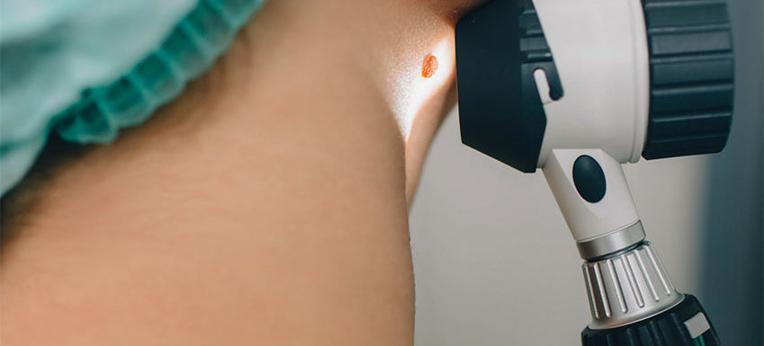
Moles are common skin growths, presenting as small, dark brown spots, sometimes flat and sometimes raised. They are formed by clusters of pigmented cells, occurring when melanocytes grow in clusters or clumps. Melanocytes are the skin cells which give us our color by producing the natural pigment known as melanin. Moles most commonly appear during childhood or adolescence and may change in appearance with time or even fade away. Although most moles are harmless, they potentially can become cancerous, developing into skin cancers like malignant melanoma. It is important that moles are assessed during your yearly skin check and that if they look suspicious they are biopsied and removed if malignant.
What Are The Signs a Mole Might Be Cancerous?
We urge our patients to follow the “ABCDE” rules of identifying possibly malignant moles. It is important to regularly self-examine your skin in addition to having a yearly dermatology check up. Take note if any of your moles fit these descriptions and if they change at all over time.
Asymmetry
If one section of your mole has a different shape, color or appearance, you should let your dermatologist know. Benign moles are usually symmetrical.
Border
Most normal moles have a clear, regularly shaped border so if yours has an irregular shape or border or is not clearly defined, it should be examined by a dermatologist.
Color
Does your mole include different colors or different shades of the same color? This is another reason to have your mole checked by a dermatologist.
Diameter
Concerning moles generally grow to a size of 6mm or larger. A larger mole should always be examined by a dermatologist, but if a smaller mole exhibits any of the other “ABCDE”s it should also be checked.
Evolving
Any mole that changes over time needs to be seen by your dermatologist. Evolutions in size, shape and color can indicate precancerous or cancerous cells.
Precancerous and cancerous moles may vary greatly in appearance. These unusual moles are called atypical moles and it is important that these suspicious moles are examined by a dermatologist.
What Factors Increase the Risk of Developing a Melanoma?
Certain factors most likely increases the risk of developing precancerous or cancerous moles. Some people have a higher risk than others. The follow may increase one’s risk:
- Large moles from birth
- Unusual or atypical moles
- Multiple moles – when you have more than 50 moles you are at a heightened risk
- Personal history or family history of precancerous or cancerous moles
When you fall into a higher risk category, it is important to be vigilant about regular self-examinations and annual dermatologist appointments.
Standard Mole Removal For Biopsy
Generally to determine what types of cells make up a mole, it needs to be removed for biopsy. Technically there is a difference between having a mole removed and having it biopsied, but the processes are very closely connected. Either way, it is normal to be concerned that mole removal may be painful and cause permanent scarring.
In a standard mole biopsy, a dermatologist uses a tool similar to a razor to shave off the mole and then uses a circular tool to remove a section or a scalpel to remove the entire mole. Once the mole is removed, generally you will receive a few stitches to help with the healing process. This procedure is done under local anesthesia and typically takes less than 10 minutes. Mole biopsy is a quick procedure, but does require administration of local anesthesia. While minimally invasive, mild tenderness and scars can occur.
DermTech Mole Biopsy
Meet DermTech, a new technique being used at APDKC. DermTech is a novel non-invasive adhesive skin biopsy collection system to test concerning pigmented lesions for cancerous cells without cutting the skin. This new and painless option eliminates needles and unnecessary scars. The sticker is simply pressed on the mole, quickly lifted off and then your skin’s RNA material is assayed for melanoma. Traditional biopsy methods relied on looking for physical changes in the cells of the mole under a microscope. The new DermTech method allows for earlier and more accurate detection that doesn’t rely on subjective visual assessment.
Protect Your Skin
While some factors in malignant moles are unavoidable, there are measures we can take to protect our skin. It is important for everyone to protect their skin from UltraViolet (UV) radiation, which can increase the risk of skin cancer. A good starting place is avoiding peak sun times to reduce intense exposure to harmful sun rays. The sun is at its strongest from 10am to 4 pm. Either avoid sun exposure during those times, wear protective clothing to cover the skin or apply sunscreen with a Sun Protective Factor (SPF) of at least 30 to help protect the skin. It’s also important to avoid sunlamps and tanning beds as they emit UV rays as well.
It is also important to examine our skin regularly and to focus on any moles or pigmentation. Use the “ABCDE” method and if there has been any change or you have reason to be worried, make an appointment for a professional skin examination.
Dr. Kaplan and the team at Adult and Pediatric Dermatology are committed to providing individualized care for all their patients. Contact our office today to schedule an appointment to discuss DermTech.

